Veterinary Pathology
Online Resource for Unique Veterinary Pathology Lesions
Vitamin D3 (Cholecalciferol) Toxicity in Rats.
Source of Images: Research Work conducted on Vitamin D3 Toxicity.
Research Plan: Animal Model: Male Wistar Albino Rats of age 42 day old, Dosing: Oral administration of Vitamin D3(Cholecalciferol) dissolved in vegetable oil @ 2 mg/kg BW single dose per day.
| | |
| Fig.1: Treated rat showing severe emaciation, cachexia and dehydration. | Fig.2: Rat from control group showing normal muscular mass at necropsy as compared to treated rats. |
| | |
| Fig.3: Necropsy of rat died due to vitamin D3 Toxicity. Note the presence of chalky white deposits on epicardial surface of heart, serosal surface of stomach & intestine. Also note the presence of pin point white deposits visible from capsular surface of kidney. | Fig.4: The kidneys from treated rats showing yellowish discoloration along with presence of pin point white deposits (Left Panel). Compare with Normal kidney from control rats (Right Panel). |
| | |
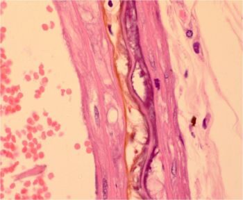
| Fig.5:Note the presence of white chalky deposits on epicardial surface of heart from rat died due to vitamin D3 toxicity (Left Panel). Heart from control Rat (Right Panel). | Fig.6:Sections of tongue from rats died due to vitamin D3 Toxicity showing purple deposits of calcification in lamima propria and connnective tissue in between muscle fibers along with lympho-mononuclear infiltration (Left Panel). Also note the deposits of calcification in tunica intima of blood vessel of tongue(Right Panel). (H&E Stain, 20X). |
| | |
| Fig.7:a)Section of stomach showing calcification of mucosa, muscularis mucosa and muscularis externa (Left Panel, H&E X 10). b) Section of stomach showing hemorrhagic gastritis and calcification of muscularis mucosae and muscularis externa (Right Panel, H&E X 20). | Fig.8:a)Section of stomach showing black deposits of mineralisation within mucosa, muscularis mucosae, muscularis externa and within walls of blood vessels. (Left Panel, Von Kossa stain X10). b) Section of large intestine showing calcification of muscularis externa layer. (Right Panel, H&EX10). |
| | |
| Fig.9: Section of kidney from rat died due to vitamin D3 toxicity showing marked coagulative necrosis and calcification (purple color deposits) within tubular epithelium and in lumen of tubules along with presence of cellular casts in lumen. H&E X40. | Fig.10: Section of kidney from rat died due to vitamin D3 toxicity showing marked coagulative necrosis and calcification (purple color deposits) within tubular epithelium and in basement membrane of tubules along with presence of cellular casts in lumen. H&E X100. |
| | |
| Fig.11:Section of kidney showing black deposits of mineralisation within cortical tubules(Left Panel, Von Kossa Stain X10). b) Section of kidney. Black deposits of mineralisation within collecting tubules in medulla. (Right Panel, Von Kossa stain X10). | Fig.12: Section of kidney showing calcification within wall of renal blood vessel. (Left Panel, Von Kossa Stain X 10). b) Section of kidney showing calcification of glomerular capsule and within glomerulus (Right Panel, Von Kossa Stain X 10). |
| | |
| Fig.13:Section of trachea showing purple deposits of calcification within cartilage and basement membrane of mucosal epithelium (Left Panel, H&E X 40). b) Section of trachea. Black deposits of calcification within cartilage and basement membrane of mucosal epithelium. (Right Panel, Von Kossa stain X 20). | Fig.14: Section of lung showing edema, hemorrhage, lympho-mononuclear infiltration and presence of giant cells. H&E X 20.
|
| | |
| Fig.15:Section of lung. Black deposits of calcium within alveolar septae. (Left Panel, Von Kossa Stain X 10). b) Section of lung. Black deposits of mineralisation within alveolar septae and bronchial mucosa. (Right Panel, Von Kossa stain X 20). | Fig.16: Section of lung showing alveolar wall thickening due to calcification(brown dirty color material)and lympho-mononuclear infiltration. (Left Panel, H&E X 40). b) Section of lung. Granular black deposits of mineralisation within alveolar septae. (Right Panel, Von Kossa stain X 100). |
| | |
| Fig.17:Section of heart. Calcification and replacement of myocardial fibers within epicardium and myocardium by fibrous tissue and lymphomononuclear cells. H&E X 20. | Fig.18: Section of heart. Calcification and replacement of myocardial fibers within epicardium and myocardium by fibrous tissue and lymphomononuclear cells.H&E X 40. |
| | |
| Fig.19: Section of heart. Marked calcification within wall of coronary blood vessel and myocardial muscle fibers. H&E X 20. | Fig.20: Section of heart. Black deposits of calcification within myocardial fibers and coronary blood vessel. Von Kossa stain X 10. |
 | 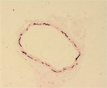 |
| Fig.21: Section of aorta showing calcification(Purple Color Deposits) within tunica media and separation and derangement of elastic lamellae. H&E X 100. | Fig.22:Section of aorta. Black deposits of mineralisation within tunica media. Von Kossa stain X 4. |
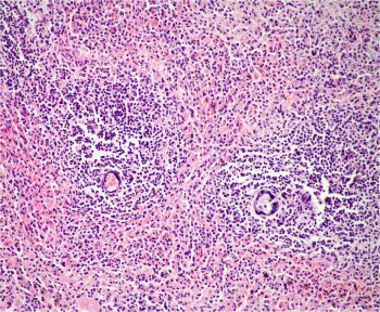 | 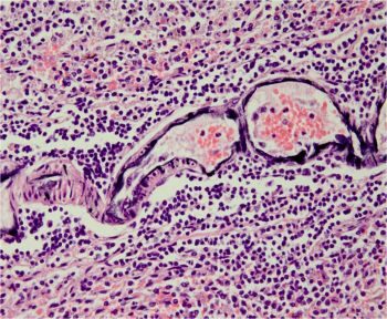 |
| Fig.23: Section of spleen showing calcification within walls of central arteries of lymphatic nodules. H&E X 20. | Fig.24: Section of spleen showing calcification of wall of central arteries of lymphatic nodules. H&E X 40. |
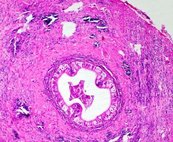 | 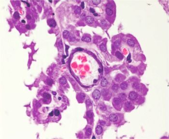 |
| Fig.25: Section of vas deferens showing calcification within muscular layer. H&E X 20. | Fig.26: Section of cerebral hemisphere. Calcification of choroid plexus blood vessel. H&E X 100. |
MECHANISM OF TOXICITY: Vitamin D3 toxicity/hypervitaminosis D ------>Causes1) Increased absorption of calcium from small intestine & Resorption of calcium from bone 2) Decreased excretion of calcium from kidney-------->hypercalcemia and hyperphosphataemia-------->calcification/mineralisation of various vital organs throughout body leading to cardiac and renal failure--------> Death of Animal.
FINDINGS:
a) Clinical Signs: The treated rats showed anorexia, difficulty in movement & respiration, diarrhoea, subnormal body temperature and progressive body weight loss. Before death, there were nervous signs like aimless running, rolling and epileptic seizures. The mortality in treated rats observed between day 8-12 of treatment.
b) Biochemical Findings: The biochemical changes observed includes, significant increase in plasma concentration of calcium, phosphorus and BUN while significant decrease in concentration of albumin and total plasma protein.
c) Gross Postmortem Findings: The gross postmortem changes observed in rats died due to toxicity includes severe emaciation, white chalky deposits on epicardial surface of heart, pin point white deposits on cortical surface of kidneys with pale yellow discoloration and diffused white deposits on serosal surface of stomach and intestine with bloody ingesta in lumen.
d) Histopathological Findings: The histopathological lesions includes generalized calcification of various organs including tongue, stomach, intestines, kidney, heart, aorta, larynx, trachea, lungs and spleen. The calcification of choroid plexus arteries, endosteal and periosteal surfaces of bones, cornea and vas deferens was also observed in some rats.
e) Ultrastructural Findings: The ultrastructural(TEM) changes in heart includes loss of mitochondrial cristae and myofibril striations. The electron micrograph of aorta showed irregular precipitations of fine needle like high electron density material.
DISCUSSION:
With unregulated increase in plasma calcium and phosphorus level in vitamin D3 toxicity, their product (calcium x phosphorus) can rise above 60 which causes mineralisation of various tissues/organs throughout body. The calcification/mineralization induced structural damage leads to decreased functional capacity of these tissues/organs. This loss of function contributes to the development of ongoing and ends stage clinical signs as well as long term signs in animals that survive. In present study, the hypercalcemia, hyperphosphatemia along with mineralisation of various organs was observed. Thus the clinical signs observed were the sequel of biochemical and histopathological changes due to vitamin D3 toxicity.
REFERENCES:
1) S G Chavhan, R S Brar, H S Banga, H S Sandhu, S Sodhi, V R Kothule, P D Gadhave and A M Kammon (2011).Clinicopathological Studies on Vitamin D3 Toxicity and Therapeutic Evaluation Aloe vera in Rats. Toxicology International 18(1): 35-43. (Another Link to Article).
***********************************************************************