Veterinary Pathology
Online Resource for Unique Veterinary Pathology Lesions
-: A Case of Intestinal Coccidiosis in Layer Poultry Birds :-
1) Case History
Layer poultry birds of age 18/19 weeks, rared on deep litter, with history of bloody diarrhea and high mortality. Environmental History: Rainy, humid environment with surrounding temperature 23 degree Celsius.
2) Gross Postmortem Lesions
| | |
| Fig.1: Note the blood mixed watery faeces oozing out from rectum/vent of dead bird at necropsy. | Fig.2: Note the blood mixed watery faeces oozing out from rectum/vent of dead bird at necropsy. |
| | |
|
Fig.3:Note the small white spots usually intermingled with rounded, bright- or dull-red spots of various sizes on the serosal surface of affected portion of the small intestine. The affected portion of small is distended twice than its normal size (ballooning) and filled with blood. The anterior and middle third portion of small intestine found prominently involved. | Fig.4:Note the small white spots usually intermingled with rounded, bright red-or dull-red spots of various sizes on the serosal surface of affected portion of the small intestine. The affected portion of small intestine is distended twice than it's normal size (ballooning) and filled with blood. The anterior and middle third portion of small intestine found prominently involved. |
| | |
|
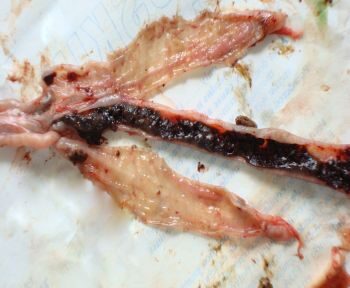
Fig.5:The affected portion of small intestine is distended twice than it's normal size (ballooning) and filled with blood. The anterior and middle third portion of small intestine found prominently involved. Ceca are apparently normal. |
Fig.6: Apparently normal gross appearance of ceca as compared to some visible portion of small intestine. |
| | |
| Fig.7: Presence of fresh or clotted blood mixed content in the lumen of small intestine. The lumen is filled with fresh or clotted blood, mucus/mucoid debris and fluid. | Fig.8: Presence of fresh or clotted blood mixed content in the lumen of small intestine. The lumen is filled with fresh or clotted blood, mucus/mucoid debris and fluid. |
| | |
| Fig.9: Note the presence of fresh or clotted blood mixed content in the lumen of small intestine. The lumen is filled with fresh or clotted blood, mucus/mucoid debris and fluid which is oozing outside when cut opened for examination. | Fig.10: Note the presence of fresh or clotted blood mixed content in the lumen of small intestine. The content is mixed with fresh or clotted blood and the mucous coat of intestine is mottled due to multiple petechial or larger haemorrhages. |
| | |
| Fig.11: Formation of Cores in the lumen of small intestine. Cores are accumulations of clotted blood, tissue debris and oocysts, in the lumen detached from affected mucosa found in later stages. Sometimes may be found in birds surviving the acute stage. | Fig.12: Core recovered from lumen of small intestine of affected bird at necropsy.
|
| | |
| Fig.13: Image showing parts/portions of intestine affected. Anterior & Middle third portion of small intestine majorly affected while ceca are apparently normal. | Fig.14: Image showing parts/portions of intestine affected. Anterior & Middle third portion of small intestine majorly affected while ceca are apparently normal. |
 |  |
|
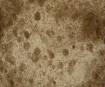
Fig.15: Image showing parts/portions of intestine affected and apparently normal appearance of ceca. | Fig.16: Note the presence of numerous unsporulated coccidial oocysts in the intestinal content/intestinal scrapings from affected birds at necropsy. (10X under low illumination). |
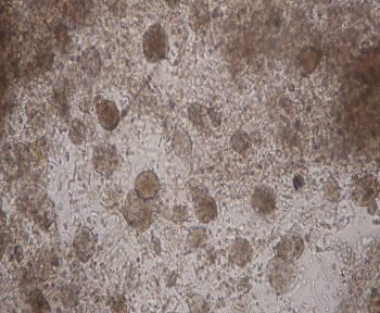 | 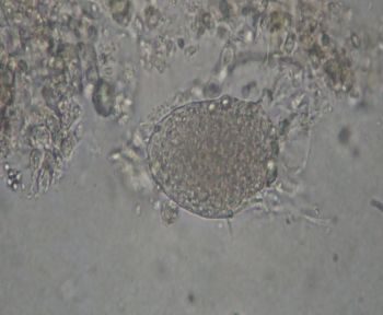 |
| Fig.17: Note the presence of numerous unsporulated coccidial oocysts in the intestinal content/intestinal scrapings from affected birds at necropsy. (10X under low illumination). | Fig.18: Unsporulated coccidial oocyst in the intestinal content/intestinal scrapings from affected birds at necropsy. (40X under low illumination). |
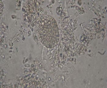 | 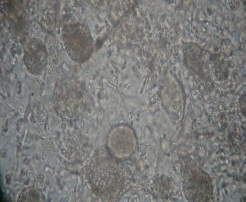 |
| Fig.19: Unsporulated coccidial oocyst in the intestinal content/intestinal scrapings from affected birds at necropsy. (40X under low illumination). | Fig.20: Note the presence of numerous unsporulated coccidial oocysts in the intestinal content/intestinal scrapings from affected birds at necropsy. (40X under low illumination). |
3) Case Discussion
The outbreak has been controlled successfully after administration of Amprolium and Sulfa Drugs. The evidences like case history, lesions at necropsy and presence of numerous coccidial oocysts in intestinal content/intestinal scrapings confirm the intestinal form of coccidiosis. Location of lesions in small intestine, age of birds, high mortality and severity of lesions(pathogenecity) suggests most probably the causative agent is Eimeria necatrix. However, the possible involvement of other Eimeria species (E.maxima, E. mitis, E. brunetti, E acervulina, E praecox etc.) cannot be denied.
